43 the human heart and its labels
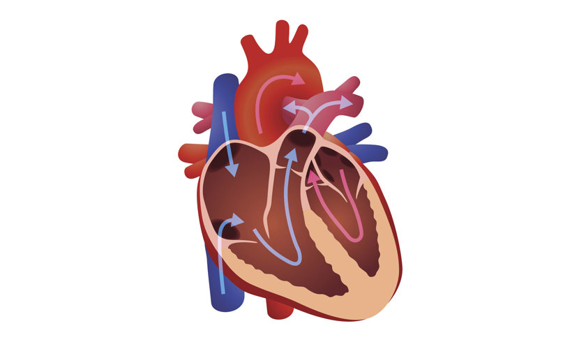
Human heart: Anatomy, function & facts | Live Science The human heart has four chambers: two upper chambers (the atria) and two lower ones (the ventricles), according to the National Institutes of Health. The right atrium and right ventricle together... A Labeled Diagram of the Human Heart You Really Need to See The human heart, comprises four chambers: right atrium, left atrium, right ventricle and left ventricle. The two upper chambers are called the left and the right atria, and the two lower chambers are known as the left and the right ventricles. The two atria and ventricles are separated from each other by a muscle wall called 'septum'.
File:Diagram of the human heart (cropped).svg - Wikipedia Diagram of the human heart, created by Wapcaplet in Sodipodi. Cropped by Yaddah to remove white space ... Add Inferior vena cava and pericardium labels: 18:08, 14 August 2018: 656 × 631 (209 KB) Jmarchn: Add pericardium. Add papillary muscles and chordae tendinae. Add cardiac skeleton. Inferior vena cava more wide.
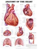
The human heart and its labels
Carefully study the diagram of the human heart with labels I II III and ... Select the option which gives correct identification and its main function. Page 36 of 53 (a) (I) Pulmonary artery: Carry oxygenated blood from heart to lungs (b) (II) Pulmonary veins: Carry oxygenated blood from lungs to the heart (c) (III) Aorta: Carry deoxygenated blood from heart to lungs (d) (IV) Vena Cava: Carry oxygenated blood from body ... Heart - Wikipedia The human heart is situated in the mediastinum, at the level of thoracic vertebrae T5-T8.A double-membraned sac called the pericardium surrounds the heart and attaches to the mediastinum. The back surface of the heart lies near the vertebral column, and the front surface sits behind the sternum and rib cartilages. The upper part of the heart is the attachment point for several large blood ... Human Heart - Anatomy, Functions and Facts about Heart - BYJUS The human heart is divided into four chambers, namely two ventricles and two atria. The ventricles are the chambers that pump blood and atrium are the chambers that receive the blood. Among which, the right atrium and ventricle make up the "right portion of the heart", and the left atrium and ventricle make up the "left portion of the heart." 5.
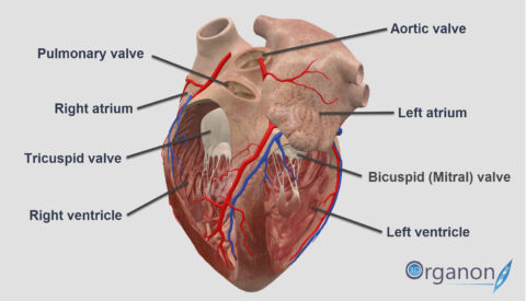
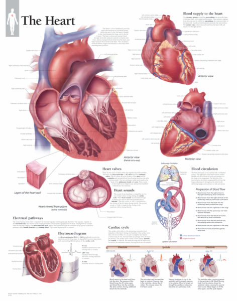
The human heart and its labels. Heart Diagram with Labels and Detailed Explanation - BYJUS Diagram of Heart. The human heart is the most crucial organ of the human body. It pumps blood from the heart to different parts of the body and back to the heart. The most common heart attack symptoms or warning signs are chest pain, breathlessness, nausea, sweating etc. The diagram of heart is beneficial for Class 10 and 12 and is frequently ... The 18 parts of the human heart, and their functions The 18 parts of the human heart and how they work 1. Myocardium 2. Endocardium 3. Pericardium 4. Right Auricle 5. Right ventricle 6. Tricuspid valve 7. Pulmonary valve 8. Left Auricle 9. Left ventricle 10. Mitral valve 11. Aortic valve 12. Tendon cords 13. Papillary muscles 14. Sinus node 15. Atrioventricular node 16. Atrioventricular fascicule 17. Anatomy of a Human Heart - U of M Health Located between the lungs in the middle of the chest, the heart pumps blood through the network of arteries and veins known as the cardiovascular system. It pushes blood to the body's organs, tissues and cells. Blood delivers oxygen and nutrients to every cell and removes the carbon dioxide and other waste products made by those cells. PDF Analyzing the Human Heart - Beyond the Classroom working. Its job is to pump blood to the lungs and to all of the body tissues. In this activity you will use a diagram of the heart to analyze the way in which the heart works. l. Using the following word list, label the various parts of the heart on the diagram. Right ventricle Left venfficle Upper vena cava Lower vena cava Aorta
Heart: illustrated anatomy - e-Anatomy - IMAIOS Anatomical structures were labelled according to the actual Terminologia Anatomica. Anatomy of the human heart and coronaries: how to visualize anatomic structures This tool provides access to several medical illustrations, allowing the user to interactively discover heart anatomy. File:Diagram of the human heart (no labels).svg File:Diagram of the human heart (no labels).svg. From Wikimedia Commons, the free media repository. File. File history. File usage on Commons. Metadata. Size of this PNG preview of this SVG file: 498 × 599 pixels. Other resolutions: 199 × 240 pixels | 399 × 480 pixels | 639 × 768 pixels | 851 × 1,024 pixels | 1,703 × 2,048 pixels | 533 × ... Labelling the heart — Science Learning Hub Labelling the heart — Science Learning Hub Labelling the heart Add to collection The heart is a muscular organ that pumps blood through the blood vessels of the circulatory system. Blood transports oxygen and nutrients to the body. It is also involved in the removal of metabolic wastes. Topics Concepts Citizen science Teacher PLD Glossary Sign in Heart Diagram - 15+ Free Printable Word, Excel, EPS, PSD Template ... Teachers and students use the heart diagram, in biological science, to study the structure and functions of a human being's heart. ... Label The Parts Of The Heart. depts.washington.edu | Having the heart diagram for studies or for scientific purpose has been made easy through this template. It shows a heart picture with all its parts labeled ...
A Look at the Human Heart - ACLS The human heart is an incredible organ that plays a vital role in how your body works. It works like a pump which pushes life-giving blood through your body. The heart is the center of the circulatory system which feeds oxygenated blood, deoxygenated blood and nutrients to vital organs and muscle tissue throughout your body; it also helps to ... 147 Heart Anatomy With Labels Premium High Res Photos - Getty Images Browse 147 heart anatomy with labels stock photos and images available, or start a new search to explore more stock photos and images. of 3. NEXT. How the Heart Works - The Heart | NHLBI, NIH - National Institutes of ... The Heart. The heart is an organ about the size of your fist that pumps blood through your body. It is made up of multiple layers of tissue. Your heart is at the center of your circulatory system. This system is a network of blood vessels, such as arteries, veins, and capillaries, that carries blood to and from all areas of your body. Human Heart Photos and Premium High Res Pictures - Getty Images Browse 21,069 human heart stock photos and images available, or search for human heart illustration or human heart icon to find more great stock photos and pictures. Related searches: human heart illustration. human heart icon. human heart vector.
Normal Anatomy of the Human Heart Giclee Print by Nucleus Medical Art - at AllPosters.com.au
heart | Structure, Function, Diagram, Anatomy, & Facts heart, organ that serves as a pump to circulate the blood. It may be a straight tube, as in spiders and annelid worms, or a somewhat more elaborate structure with one or more receiving chambers (atria) and a main pumping chamber (ventricle), as in mollusks. In fishes the heart is a folded tube, with three or four enlarged areas that correspond to the chambers in the mammalian heart. In animals ...
Sampoerna Wallpaper: The Heart Diagram Labeled
Diagram of the human heart royalty-free images - Shutterstock 14,791 diagram of the human heart stock photos, vectors, and illustrations are available royalty-free. See diagram of the human heart stock video clips Image type Orientation Color People Artists Sort by Popular Anatomy Healthcare and Medical Icons and Graphics Diseases, Viruses, and Disorders heart medicine organ hemodynamics circulatory system

Pokemon Cosplay: Kawaii Pokemon Pikachu Cosplay Couple
13 parts of the human heart (and its functions) - LORECENTRAL Parts of the heart and its functions 1. Left atrium 2. Mitral Valve 3. Left Ventricle 4. Aortic sigmoid valve Right atrium 6. Tricuspid valve 7. Right ventricle 8. Pulmonary sigmoid valve 9. Atrial septal defect Interventricular partition 11. The sinus or sinoatrial node 12. Atrioventricular or Aschoff-Tawara nodule 13. Hiscules and Purkinje fibers

New Photos in Anatomy of human body organs
Label the heart — Science Learning Hub In this interactive, you can label parts of the human heart. Drag and drop the text labels onto the boxes next to the diagram. Selecting or hovering over a box will highlight each area in the diagram. aorta vena cava left atrium semilunar valve pulmonary vein right atrium right ventricle pulmonary artery left ventricle Download Exercise Tweet
Our World | What is the heart?
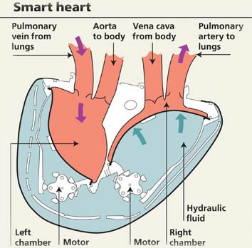
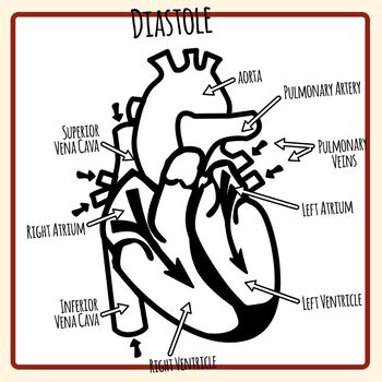
A Diagram of the Heart and Its Functioning Explained in Detail The heart blood flow diagram (flowchart) given below will help you to understand the pathway of blood through the heart.Initial five points denotes impure or deoxygenated blood and the last five points denotes pure or oxygenated blood. 1.Different Parts of the Body ↓ 2.Major Veins ↓ 3.Right Atrium ↓ 4.Right Ventricle ↓ 5.Pulmonary Artery ↓ 6.Lungs
How the human heart did become associated with love? | 3D ORGANON
Human Heart - Diagram and Anatomy of the Heart - Innerbody The heart contains 4 chambers: the right atrium, left atrium, right ventricle, and left ventricle. The atria are smaller than the ventricles and have thinner, less muscular walls than the ventricles. The atria act as receiving chambers for blood, so they are connected to the veins that carry blood to the heart.

Parts Of Heart Diagram Stock Illustration - Download Image Now - iStock
Human Heart (Anatomy): Diagram, Function, Chambers, Location in ... - WebMD The heart is a muscular organ about the size of a fist, located just behind and slightly left of the breastbone. The heart pumps blood through the network of arteries and veins called the...
Human Heart - Label the Diagram Anatomy Clip Art Set Commercial Use
How to Draw a Human Heart: 11 Steps (with Pictures) - wikiHow Method 1Sketching the Heart. 1. Draw the lower half of an acorn shape so it's tilted to the left. Use your pen or pencil to start drawing the main part of the heart. This should look like an open-ended acorn that's missing its cap. Draw the shape so it's tilted about 120 degrees to the left.

Simplified Heart Labeled Decal | Shop Fathead Anatomical Images Graphics
The Anatomy of the Heart, Its Structures, and Functions - ThoughtCo The heart is the organ that helps supply blood and oxygen to all parts of the body. It is divided by a partition (or septum) into two halves. The halves are, in turn, divided into four chambers. The heart is situated within the chest cavity and surrounded by a fluid-filled sac called the pericardium. This amazing muscle produces electrical ...

Parts Of The Human Heart | Science Trends The parts of the human heart can be broken down into four chambers, muscular walls, vessels, and a conductive system. The two upper chambers are called the atria, with lower parts called ventricles. These all work together to make up the vital function of your heart. Everybody knows that the human heart is the essential organ in our bodies.

Congestive Heart Failure: The Essence of Heart Failure Course | CEUfast Nursing Continuing Education
How the Heart Works: Diagram, Anatomy, Blood Flow - MedicineNet The heart is an amazing organ. It starts beating about 22 days after conception and continuously pumps oxygenated red blood cells and nutrient-rich blood and other compounds like platelets throughout your body to sustain the life of your organs.; Its pumping power also pushes blood through organs like the lungs to remove waste products like CO2.; This fist-sized powerhouse beats (expands and ...

Human Heart Diagram Educational Posters EP126
Diagram of Human Heart and Blood Circulation in It Exterior of the Human Heart A heart diagram labeled will provide plenty of information about the structure of your heart, including the wall of your heart. The wall of the heart has three different layers, such as the Myocardium, the Epicardium, and the Endocardium. Here's more about these three layers. Epicardium
The Heart | Scientific Publishing
Human Heart - Anatomy, Functions and Facts about Heart - BYJUS The human heart is divided into four chambers, namely two ventricles and two atria. The ventricles are the chambers that pump blood and atrium are the chambers that receive the blood. Among which, the right atrium and ventricle make up the "right portion of the heart", and the left atrium and ventricle make up the "left portion of the heart." 5.

30 Human Heart With Label - Labels Design Ideas 2020
Heart - Wikipedia The human heart is situated in the mediastinum, at the level of thoracic vertebrae T5-T8.A double-membraned sac called the pericardium surrounds the heart and attaches to the mediastinum. The back surface of the heart lies near the vertebral column, and the front surface sits behind the sternum and rib cartilages. The upper part of the heart is the attachment point for several large blood ...

Human Heart Pictures with Labels Best Of File Diagram Of the Human Heart Hug Wikimedia Mons ...
Carefully study the diagram of the human heart with labels I II III and ... Select the option which gives correct identification and its main function. Page 36 of 53 (a) (I) Pulmonary artery: Carry oxygenated blood from heart to lungs (b) (II) Pulmonary veins: Carry oxygenated blood from lungs to the heart (c) (III) Aorta: Carry deoxygenated blood from heart to lungs (d) (IV) Vena Cava: Carry oxygenated blood from body ...

Your New Puppy Author Cindy Moore, cindy@k9web.com Copyright 1992-99. Table of Contents ...

Business Diary: October 2011
Post a Comment for "43 the human heart and its labels"