39 colon diagram with labels
Intestines (Anatomy): Picture, Function, Location, Conditions The intestines include the small intestine, large intestine, and rectum. The small intestine (small bowel) is about 20 feet long and about an inch in diameter. Its job is to absorb most of the ... Illustration Picture of Anatomy - Colon - eMedicineHealth The colon is the largest part of the large intestine, extending from the cecum to the rectum. It is 5 feet long and its function is to reabsorb water from digested food and concentrate solid waste material, known as stool. The colon is made of several sections. The ascending colon travels up the right side of the abdomen.
Labeled Diagram Of The Digestive System Stock Photos, Pictures ... Labeled human anatomy diagram of male shoulder, biceps, arm, and chest muscles frontal anterior view on a white background. Chicken anatomy vector illustration. Labeled biological inner... Chicken anatomy vector illustration. Labeled biological inner organs scheme. Zoological graphic with birds bones, digestive system and inside structure.

Colon diagram with labels
Generate music with an RNN | TensorFlow Core May 27, 2022 · In this way, the model will be trained to predict the next note in a sequence. You can find a diagram explaining this process (and more details) in Text classification with an RNN. You can use the handy window function with size seq_length to create the features and labels in this format. Use the Create Diagram from Data wizard - support.microsoft.com You can use the Create Diagram from Data wizard to create a detailed, polished Visio flowchart from an Excel workbook. Follow the steps in the wizard and use this help information if you have questions in each step. For more information about Data Visualizer, see Create a Data Visualizer diagram. Colon: Anatomy, histology, composition, function | Kenhub The colon forms part of the large intestine and extends between the caecum and the rectum. It is about 1.5 meters in length and consists of four parts: ascending transverse descending sigmoid colon You can recognize it easily through several distinct morphological features like semilunar folds and pouches called haustra.
Colon diagram with labels. PDF Digestive System Diagram Label these: #1 (Urine/pee pee) #2 (Solid Waste/poop) Hydrochloric Mechanical Digestion Chemical Digestion Saliva Acid Pepsin Bile Lipase Stomach Small Intestine Enzymes from Liver and Pancreas Large Intestine (Transverse Colon) Descending ColonCirculatory System Kidneys #1 #2 Water and Vitamins Nutrients The Digestive System Blog - Create a sequence diagram This works similar to release numbering, where each lifeline adds another point. Add the message number and a colon at the start of the message label. E.g. 1.3: searchByItem(itemName) Open this sequence diagram in the diagrams.net viewer. Frame labels for sequence fragments. The type of sequence fragment is written in the top left of the frame ... PDF ANATOMIC DRAWINGS OF THE DIGESTIVE SYSTEM Esophageal sphincter Liver ... Colon (C18._).0 Cecum.1 Appendix.2 Ascending.3 Hepatic flex..4 Transverse.5 Splenic flex..6 Descending.7 Sigmoid.8 Overlapping.9 Colon, NOS Yes Yes Yes Yes Yes Yes Yes Yes Yes Yes Yes Yes Yes Yes Yes Yes Yes Yes Yes Yes Yes Yes Yes Yes Yes Yes Yes Yes Yes Yes Yes Yes Yes Yes Yes Yes Yes Yes Yes Yes Yes Yes Yes Yes Yes Yes Yes Yes Yes Yes Yes ... Abdomen and digestive system anatomy: diagrams labeled Full labeled anatomical diagrams - Anatomy of the abdomen and digestive system: these general diagrams show the digestive system, with the major human anatomical structures labeled (mouth, tongue, oral cavity, teeth, buccal glands, throat, pharynx, oesophagus, stomach, small intestine, large intestine, liver, gall bladder and pancreas).
Colon Diagram Stock Illustrations - 3,304 Colon Diagram ... - Dreamstime Download 3,304 Colon Diagram Stock Illustrations, Vectors & Clipart for FREE or amazingly low rates! New users enjoy 60% OFF. 187,781,852 stock photos online. ... Human Anatomy. Labeled Diagram. Human colon. Colon - lymphatic drainage. Pathways of lymphatic drainage of the colon. Anatomy of the Colon, Rectum and Anus. Digestive system, large ... Sigmoid colon - Definition, Anatomy and Function | Kenhub Sigmoid colon - ventral view. The gastrointestinal system is divided into the foregut, midgut and hindgut.The foregut stretches from the oesophagus to the major duodenal papilla, the midgut from the major duodenal papilla to two thirds of the transverse colon, and the hindgut from this point to the pectinate line of the rectum.. Neurovasculature. The hindgut gets its blood supply from the ... Colon Picture Labeling Flashcards | Quizlet Colon Picture Labeling STUDY Flashcards Learn Write Spell Test PLAY Match Gravity Created by XertziePLUS Terms in this set (11) rectum 10 external anal sphincter 12 teniae coli 14 cecum 8 vermiform appendix 9 transverse colon 2 ascending colon 5 ileum 6 ileocecal valve 7 sigmoid colon 13 descending colon 16 YOU MIGHT ALSO LIKE... Cecum Histology Slide with Labeled Image and Diagram The tunica muscular layer of the provided cecum labeled image shows two distinct smooth muscle layers - inner longitudinal or oblique bundles and outer wavy bundles. Again, the cecum images show some elastic fibers in this layer. In addition, the cecum labeled image shows a thin and loose connective tissue layer with numerous blood vessels.
Labeled Diagram of the Human Kidney - Bodytomy The renal medulla comprises a set of 8-18 conical structures called renal pyramids that are surrounded by the cortex. Portions of the cortex between two adjacent pyramids are termed as renal columns. Spread in these pyramids and the cortex, are the functional units callednephrons. The actual filtration of blood occurs in the nephrons. Picture of the Human Colon Anatomy & Common Colon Conditions - WebMD The ileum (last part of the small intestine) connects to the cecum (first part of the colon) in the lower right abdomen. The rest of the colon is divided into four parts: • The ascending colon... 40 Colon diagram Vector Images, Colon diagram Illustrations - Depositphotos 40 Colon diagram Stock Vector Images, Royalty-free Colon diagram Drawings & Illustrations. VectorMine Crohns disease vector illustration. Labeled diagram with diagnosis. VectorMine Ulcerative colitis vector illustration. Labeled anatomical infographic. News Archives | Hollywood.com Travel through time by exploring Hollywood.com's entertainment news archives, with 30+ years of entertainment news content.
Reproductive Health and Fetal: May 2011
Histology | Colon This diagram illustrates the 4 basic layers of the colon. The inner pink layer is the mucosa, the yellow layer beneath the mucosa is called the submucosa, while the red layer is the muscular layer (muscularis) and the 4 th layer is called the serosa or adventitia. Courtesy Ashley Davidoff MD 32338 Ultrasound of normal large bowel

The function of the small intestine in the human digestive system | Science online
Healthy Eating Plate vs. USDA’s MyPlate | The Nutrition ... The Healthy Eating Plate encourages consumers to choose fish, poultry, beans or nuts, protein sources that contain other healthful nutrients. It encourages them to limit red meat and avoid processed meat, since eating even small quantities of these foods on a regular basis raises the risk of heart disease, diabetes, colon cancer, and weight gain.

AMICUS Illustration of amicus,anatomy,bowel,normal,liver,stomach,transverse,colon,intestine ...
Understanding the Human Stomach Anatomy With Labeled Diagrams Given below is a labeled diagram of the stomach to help you understand stomach anatomy. The stomach is divided into four parts. These include: Cardia Fundus Body Pylorus Cardia refers to the section of the stomach that is located around the cardiac orifice. The lower esophageal sphincter lies at the junction where the esophagus meets the stomach.
Print A&P CH23 Digestive System flashcards | Easy Notecards
Colonoscopy Measurements (cm) from Anal Verge | SEER Training Types of Surgery: Colon; Types of Surgery: Rectum; Radiation Therapy; Commonly Used Drugs; For hands-on exercises, please go to SEER*Educate. Resources. Archived Modules. Updates. Acknowledgements. Colonoscopy Measurements (cm) from Anal Verge. Return to Anatomy of Colon and Rectum. Follow SEER. Contact Information.

VIP LASER CLINIC - MAURITIUS - Colon Hydrotherapy
Female anatomy diagram Stock Photos and Images - Alamy Find the perfect Female anatomy diagram stock photo. Huge collection, amazing choice, 100+ million high quality, affordable RF and RM images. No need to register, buy now!
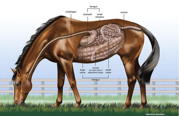
Understanding the Horse Digestive System - SmartPak
Simple Guide on Creating Flowchart for Switch Statement Dec 15, 2021 · Each case is then followed by different case labels that always end with a colon (:). The value A, B, and n are case labels that are used for identifying each case individually. Be sure that none of the case labels is the same and each is named according to the preference of execution. For example, two cases have been labeled X.

Colon Facts for Kids
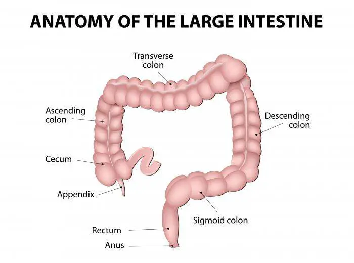
Colon Anatomy (with Small Intestine Label) - NCI Visuals Online 720x602. View. Download. Title: Colon Anatomy (with Small Intestine Label) Description: Drawing shows the cecum, ascending colon, transverse colon, descending colon, sigmoid colon, rectum, and anal canal. Also shown is the small intestine. The cecum connects the small intestine to the colon.
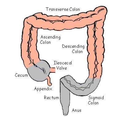
Diagram of The Colon
female pelvis diagram labeled And Pelvis Labeled Structures Include Large Bowel Colon Diagrams Female medicinebtg.com. colon pelvis bowel medicinebtg urinary excretory. Male Reproductive System Model Labeled - Google Search | Female . reproductive urethra gland organs physiology prostate scrotum anato biz. Ilium (bone). Male reproductive system model ...
colon figure | Anatomy System - Human Body Anatomy diagram and chart images
Digestive System with Labels Focusing on the Colon, Rectum, and Anus Black and white illustration of the digestive system with colon, rectum, and anus highlighted and parts labeled: esophagus, stomach, liver, gallbladder, duodenum ...

colon_diagram – Digestive Health Centre
Diagram Of The Respiratory System With Labels Pictures, Images ... - iStock A medical diagram showing the lobes of the lungs (organ of the respiratory system) with text labels. Human Lungs Diagram Cross section & anterior view of the human lungs. The digestive system The human digestive system medical illustration with internal organs Human Respiratory System Lungs Label Design Anatomy

09/09/15 ~ Nursing
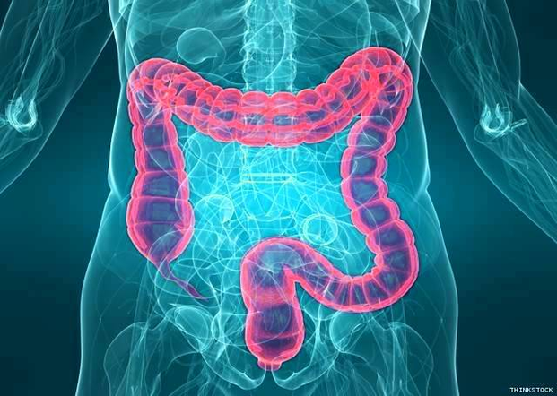
The Colon - Ascending - Transverse - Descending - TeachMeAnatomy The colon (large intestine) is the distal part of the gastrointestinal tract, extending from the cecum to the anal canal. It receives digested food from the small intestine, from which it absorbs water and electrolytes to form faeces. Anatomically, the colon can be divided into four parts - ascending, transverse, descending and sigmoid.

Anything & Everything Equine :): Digestive System of a Horse
Large Intestine Anatomy, Parts, Diagram & Major Function - Study.com Draw a diagram of the large intestine and label all parts. Updated: 07/22/2021 Table of Contents. What is a Large Intestine? ... The three parts of the colon are the ascending colon, ...
![Untitled Document [siera104.com]](http://siera104.com/school/biology/chordates/CatSkeleton.png)
Untitled Document [siera104.com]
Parts of the colon | Understanding Your Colon or Rectal Surgery The colon (large intestine) is a long, muscular tube about four to five feet long. The colon removes water and nutrients from partially digested food. Then it turns the rest into stool (waste). The stool goes through the rectum and then leaves the body through the anus. Parts of the colon. Cecum: This is the beginning of the colon. It is ...

# 52 Human alimentary canal | Biology Notes for IGCSE 2014
Colon (Large Intestine): Anatomy, Function, Structure Sigmoid colon: The S-shaped connection between the last part of the colon and the rectum, located on the bottom left side of the abdomen is called the sigmoid colon. 2 Size and Length This organ is called the large intestine because of the diameter (width) of the intestine; it is much wider than the small intestine, but also much shorter.
Colon diagram | Healthiack
Digestive organs: Diagram, stomach, intestines, and more Summary. The digestive organs in the abdomen work together to absorb nutrients and move food through the digestion process. They include the stomach, gallbladder, liver, pancreas, intestines, and ...
Post a Comment for "39 colon diagram with labels"